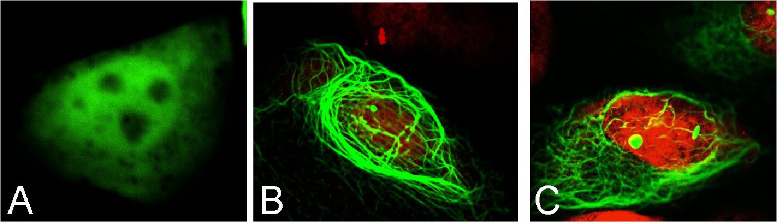
Fig. 2. EGFP was localized in a diffuse pattern both in the nucleus and the cytoplasm (A), while AIRE-EGFP (B) and CARD-EGFP (C) were localized heavily on the cytoskeletal network and nuclear speckles. Green channel shows EGFP localization, while the red channel shows propidium iodide staining of the nucleus.